

When the upper airway limits therapy
MI-E effectiveness depends on enough air reaching the lungs during insufflation and on a high expiratory flow being generated during exsufflation. In patients with amyotrophic lateral sclerosis (ALS) and bulbar dysfunction, upper airway responses can compromise these prerequisites. [2, 7]
Impaired coordination of the laryngeal and pharyngeal musculature can cause the upper airway to narrow or transiently close during the pressure changes. During insufflation in particular, paradoxical adduction of the glottis or supraglottic structures can occur. During the subsequent exsufflation, the negative pressure can promote pharyngeal narrowing or, where vulnerability exists, favour upper airway collapse. [1, 2]
This can impair airflow during MI-E, making cough augmentation less effective. Higher insufflation and exsufflation pressures often increase peak flow, but, especially with pronounced respiratory muscle weakness, they can increasingly lead to upper airway closure. [3]
A high or rising peak cough flow (PCF) does not, however, automatically mean that the upper airway is freely patent. High peak flow values can still be measured even with increasing obstruction. Alongside PCF, the pressure and flow curves, and, where available, the volumes, as well as the actual secretion mobilisation achieved, should therefore be taken into account. [2, 3]
Possible upper airway responses should accordingly be recognised and MI-E settings adjusted individually. The following sections explain which clinical and device-derived cues are relevant, what options exist for optimising therapy, and under what conditions the effectiveness of the technique can remain limited.
Two distinct obstruction patterns in bulbar dysfunction
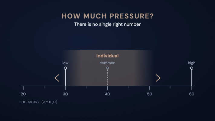
A 2022 prospective study examined 69 patients with ALS to identify which abnormalities occur during MI-E and how these relate to the respective pattern of bulbar impairment. Pressure levels of ±20, ±30, ±40 and ±50 cmH₂O were used, and a total of 351 pressure and flow recordings were analysed. [1]
Two distinct upper airway obstruction patterns emerged:
- Obstruction during insufflation: around half of the patients showed obstruction during the positive pressure phase. This pattern was associated in particular with upper motor neuron bulbar impairment. [1]
- Collapse during exsufflation: in 18 patients (26%), upper airway collapse was observed during the negative pressure phase. This pattern was associated with lower motor neuron bulbar impairment. [1]
The results show that the upper airway can respond differently to the pressure phases of MI-E, and that these responses are associated with the respective pattern of bulbar impairment. A blanket increase in insufflation or exsufflation pressure is therefore not necessarily the right approach. Analysing the pressure and flow curves can instead help tailor the settings individually.
After corresponding adjustment of the MI-E settings, cough augmentation judged effective per the study criteria was achieved in 68 of 69 patients (98.6%). The observed obstruction patterns therefore did not generally preclude effective MI-E, but made targeted adjustment of therapy parameters necessary. [1]
The Norris Bulbar Score, a clinical rating scale for bulbar function covering, among other things, speech and swallowing, was also examined. In the statistical analysis, this score was the only parameter studied that predicted the absence of abnormal pressure and flow curves. [1]
Pressure and flow curves: what to look for
Pressure and flow curves give clues to how the upper airway responds to the pressure changes during insufflation and exsufflation. It is important always to view pressure and flow together across the entire respective phase, not just the displayed peak cough flow. Where available, inspiratory and expiratory volumes can also contribute to the assessment. [2, 4]
- During insufflation: despite a positive pressure having built up, inspiratory flow is conspicuously low or flattens early toward near zero. This can indicate narrowing or closure of the upper airway.
- During exsufflation: despite negative pressure being applied, expiratory flow drops sharply early on or shows a conspicuously flat course near zero. Such a pattern can suggest dynamic upper airway collapse.
- Interpret peak flow with caution: a high peak flow value displayed at the start of exsufflation does not rule out obstruction. The pressure change can also generate a flow component from decompression of the tubing circuit that does not correspond to an effective cough. [2]
Flattening alone, however, does not prove obstruction. Before interpretation, factors such as mask leaks, where the curve falls within the breath phase, the set times and possible normal glottic movements should be taken into account. Curve findings must always be cross-checked against actual secretion mobilisation and clinical observation. [2, 4]
From curve analysis to individual therapy adjustment
Where there are indications of obstruction, adjustment of the MI-E parameters depends above all on which pressure phase shows impaired airflow. A blanket pressure increase is then often not the right approach. Instead, settings should be checked stepwise, guided by the patient's response. [1, 4, 5]
If obstruction during insufflation is suspected, it can be useful to reduce the positive insufflation pressure and the inspiratory flow. A slower rise in pressure and flow can improve the laryngeal response. If this lowers the inspiratory volume, an adjusted, possibly longer, insufflation time can help. In suitable, cooperative patients, patient-triggered insufflation can also support synchronisation. [4, 5]
Where there are indications of collapse during exsufflation, the negative exsufflation pressure in particular should be reviewed: a smaller magnitude of negative pressure can reduce the collapse. The exsufflation time should also be assessed against the flow trace and adjusted if needed. If abnormalities occur in both phases, both pressure phases and the inspiratory flow may need to be changed. [1, 4]
After each adjustment, the pressure and flow curves should be reassessed and checked against achievable volumes, tolerability and actual secretion clearance. No single pressure scheme valid for all patients can be derived from the patterns described. Titration belongs in the hands of appropriately qualified clinical staff. If findings remain unclear or cough augmentation remains inadequate, supplementary endoscopic assessment can be useful. [2, 4, 5]
Endoscopy: when a supplementary assessment is useful
Analysing the pressure and flow curves during MI-E can provide initial clues to upper airway obstruction. It does not, however, show which anatomical structures are responsible for the narrowing. This is where a supplementary endoscopic examination can be useful.
Flexible transnasal laryngoscopy allows direct observation of how the upper airway responds to the positive and negative pressure phases of MI-E. For example, paradoxical glottic adduction or closure of supraglottic structures can be visualised this way.
Particularly where obstruction patterns remain unclear or cough augmentation stays inadequate despite adjusted MI-E settings, endoscopy can help clarify the underlying causes and tailor therapy more precisely.
Endoscopy can thus be a useful supplement to MI-E assessment, especially where the pressure and flow curves do not allow clear conclusions about possible obstruction. Its value for assessing the upper airway was already described in 2019, though the lack of a validated examination protocol at the time was also noted. A more recent 2024 review underscores in particular the value of direct endoscopic assessment combined with pressure and flow curve analysis for more targeted adjustment of MI-E settings. Routine endoscopic use in all MI-E patients cannot, however, be derived from the evidence available to date. [2, 6, 7]
When MI-E reaches its limits despite adjustment
Even with careful adjustment of MI-E settings, cough augmentation can remain inadequate in pronounced bulbar dysfunction. In this situation, not only the device settings but the entire secretion management approach should be reviewed. Alongside impaired cough function, swallowing disorders and inadequate control of oral secretions can compromise airway protection and raise aspiration risk. [8]
Depending on the clinical situation, additional measures for secretion control and clearance may be needed. Supplementary techniques such as high-frequency chest wall oscillation can, for example, support mobilisation of peripheral secretions, but they do not replace adequate clearance of secretions from the central airways. [8]
If cough augmentation remains inadequate despite individual adjustment, further therapeutic options should be assessed jointly, taking into account the clinical course, the respiratory situation, and the patient's wishes. [8]
Questions about the practical application of MI-E?
Do you care for patients with ALS and bulbar dysfunction and would like more confidence setting up MI-E or assessing pressure and flow curves?
Feel free to get in touch. We can help arrange hands-on training or put you in contact with a local partner to discuss questions on application and individual therapy adjustment together.
Frequently asked questions
Why can a cough assist device be less effective in ALS with bulbar dysfunction?
In bulbar dysfunction, impaired coordination of the laryngeal and pharyngeal musculature can cause narrowing or closure of the upper airway. This can occur during both insufflation and exsufflation and impair airflow, which can limit MI-E effectiveness. [1, 2]
Does MI-E have to be discontinued if an abnormal obstruction pattern is found?
No, not necessarily. An abnormal obstruction pattern does not automatically mean that MI-E is ineffective. Cough augmentation can often be improved through individual adjustment of therapy parameters. In a prospective study of 69 ALS patients, cough augmentation judged effective per the study criteria was achieved in 68 patients after corresponding adjustment. [1]
Why doesn't higher MI-E pressure always lead to better cough augmentation?
In bulbar dysfunction, higher insufflation pressures can promote narrowing at the level of the larynx. Stronger negative pressure during exsufflation can likewise contribute to upper airway collapse. Increasing pressure therefore does not necessarily improve secretion mobilisation and can even worsen existing obstruction. [1, 3]
Does a high peak cough flow automatically mean effective cough augmentation?
No. A high or rising peak cough flow (PCF) does not automatically mean that the upper airway is freely patent during MI-E. Measurement artefacts can also affect the displayed value. Alongside peak cough flow, the pressure and flow curves and the secretion mobilisation actually achieved should therefore be assessed. [2, 3]
When is an endoscopic examination useful for MI-E?
A supplementary endoscopic examination can be useful when pressure and flow curves show indications of upper airway obstruction whose cause remains unclear. Even with inadequate cough augmentation despite individual adjustment of MI-E settings, laryngoscopy can help visualise possible narrowing or collapse of the upper airway. [2, 6]
What should be done if MI-E remains insufficiently effective despite individual adjustment?
If cough augmentation remains inadequate, the curve traces, possible leaks, the parameters set, and the actual secretion mobilisation should first be reviewed again. Where the airway response remains unclear, supplementary endoscopy can be useful. At the same time, overall secretion management, including oral secretions and possible additional measures, should be assessed. [2, 6, 8]
How can upper airway obstruction during MI-E be recognised?
Abnormal pressure and flow curves can indicate narrowing or collapse of the upper airway. Flattened flow traces during insufflation or exsufflation can be a typical finding. Curve analysis supports the assessment but does not allow reliable anatomical localisation of the obstruction. [1, 2]
Sources
Note
This article is provided for professional information and does not replace clinical judgement. Statements on device settings are orientation only; the device's instructions for use and the assessment of the treating clinician prevail.